1. Diagnostic basis.
Gastric cancer should be diagnosed and differentiated in combination with clinical manifestations, endoscopy, histopathology and imaging examination.
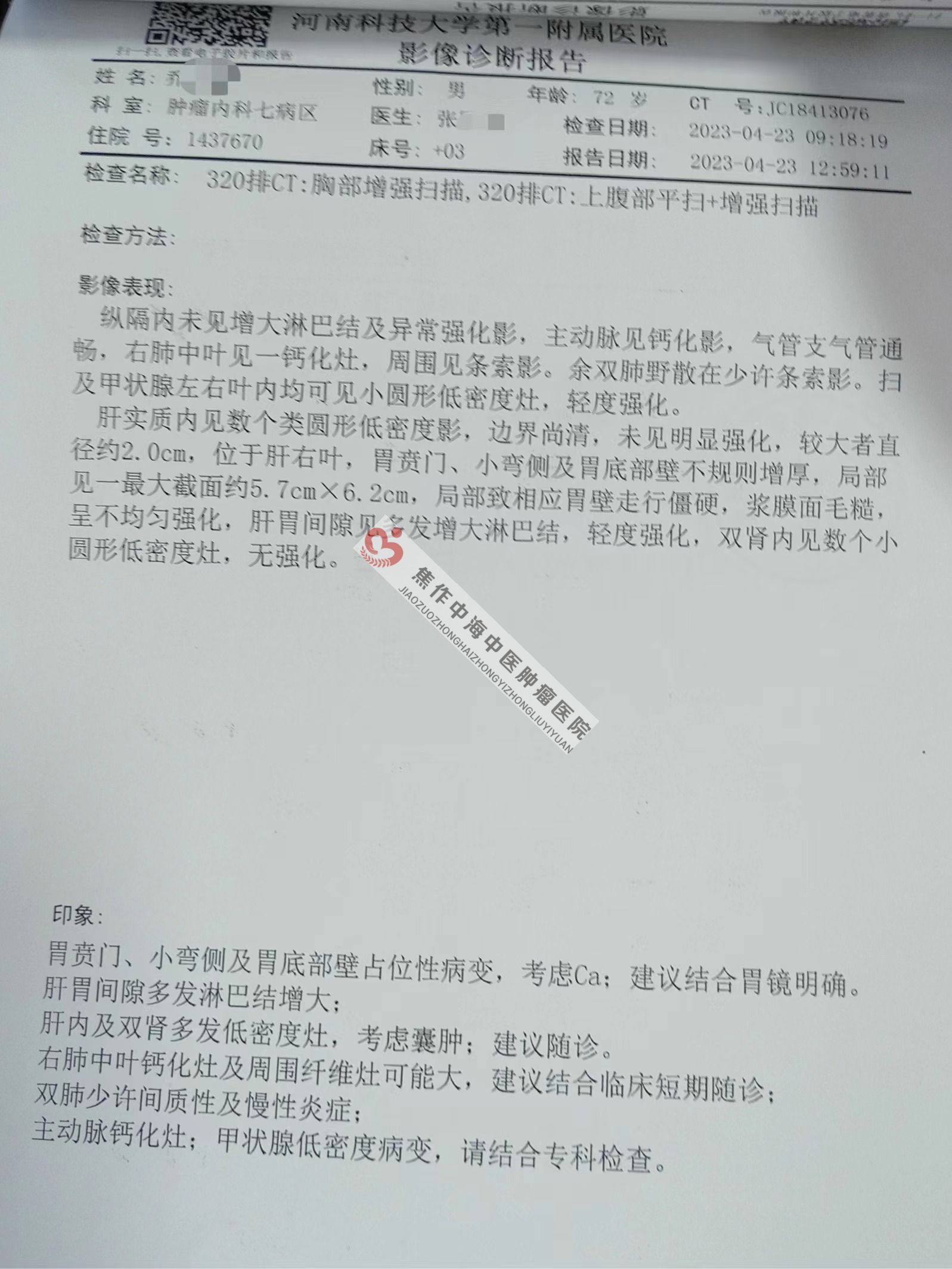
1. clinical manifestation.(1) symptoms:Early gastric cancer is often asymptomatic or has only one-some non-specific gastrointestinal symptoms. Once there are obvious symptoms, most of the patients are progressive or have complications.The common symptoms of advanced gastric cancer are. Upper abdominal discomfort or pain, loss of appetite (especially for meat), postprandial fullness, nausea or vomiting, hematemesis or black feces, dysphagia, diarrhea or constipation, fatigue, weight loss, increased abdominal circumference, anemia, low fever, etc.(2) physical signs:Early gastric cancer often has no obvious physical signs. Patients with advanced gastric cancer can touch a mass in the upper abdomen with tenderness. The left clavicle may appear when there is distant metastasis. Enlarged upper lymph nodes (Virchow lymph nodes) and left axillary lymph nodes (lrish nodules). Liver metastasis can cause hepatomegaly, jaundice and ascites. Peritoneal implant metastasis can form umbilical nodule (Sister MaryJoseph nodule), ovarian mass (Krukenberg tumor), anterior rectal mass (Blumer's shelf), ascites, abdominal distension and mobility dullness positive. Splenomegaly occurs when invading the portal vein or splenic vein. Gastric type, peristaltic wave and vibrating sound may occur in pyloric obstruction.(3) accompanied by cancer syndrome:Including recurrent superficial thrombophlebitis (Trousseau sign) and hyperpigmentation, acanthosis nigricans (pigmentation in skin folds, especially in the two axils), dermatomyositis, membranous nephropathy, microvascular hemolytic anemia and neuromyopathy.(4) complications:Including upper gastrointestinal bleeding, perforation, gastrointestinal fistula and digestive tract obstruction (cardiac, pyloric or biliary obstruction). Common postoperative complications include anastomotic fistula, vitamin B12 deficiency and so on.two。. Laboratory examination.(1) whole blood cell count: anemia is common, about 50% have iron deficiency anemia, which is caused by long-term blood loss or nutrition.Lack is caused. If combined with malignant anemia, megaloblastic anemia can be seen. Hemolytic anemia caused by microvascular disease has also been reported.Tao.(2) biochemical examination: abnormal liver function indicates that there may be liver metastasis.(3) fecal occult blood test: fecal occult blood test is often continuously positive, which is of auxiliary diagnostic significance.(4) the tumor marker CEA is elevated in 30 ~ 40% of patients with primary gastric cancer, which has a certain value in follow-up rather than screening or diagnosis. Other tumor markers (CA199, CA125, CA724) may increase in varying degrees in some patients with gastric cancer, but they have no screening or diagnostic value.3. Endoscopic examination.(1) fiberoptic gastroscopy:It is the most reliable diagnostic method to determine whether the tumor exists and its location, and to biopsy any suspicious lesions. Almost all patients with gastric cancer diagnosed by upper gastrointestinal radiography need to undergo fiberoptic gastroscopy and biopsy to confirm the diagnosis. Pigment endoscope or magnifying endoscope can be used when necessary.(2) Endoscopic ultrasound (EUS):It is very important for the clinical staging of gastric cancer before initial treatment. It is helpful to evaluate the depth of invasion (T stage), submucosal spread and perigastric lymph node metastasis (N stage), and sometimes distant dissemination signs (M stage) can be found, such as surrounding organ lesions or ascites. This examination is necessary for minimally invasive operations such as endoscopic mucosal resection (EMR) and endoscopic submucosal resection (ESD).Because the detection depth of EUS is shallow and the visibility of the sensor is limited, the accuracy of evaluating distant lymph node metastasis is not satisfactory.(3) Laparoscopy:Suitable for T3 or N1 with no metastasis (M0) in imaging examination, laparoscopic staging and peritoneal lavage should be considered for cytological examination. Positive peritoneal cytology (with visible peritoneal implantation metastasis) has a poor prognosis and is defined as M1 disease. Laparoscopic exploration plays a limited role in the evaluation of liver metastasis and perigastric lymph node metastasis.4. Imaging examination.(1) computed tomography.(CT): CT plain scan and enhanced scan have important value in evaluating the lesion extent, local lymph node metastasis and distant metastasis of gastric cancer. It has been routinely used in preoperative staging of patients with gastric cancer, and its accuracy of T staging has reached 43% ~ 82%.(2) MRI examination:MRI is one of the most important imaging methods. It is recommended for patients with CT contrast agent allergy or other suspected metastases in imaging examination. MRI is helpful in judging peritoneal metastasis and can be used as appropriate.(3) Upper gastrointestinal radiography:It is helpful to judge the location, scope and functional state of primary gastric lesions, especially air-barium double contrast radiography is one of the common imaging methods in the diagnosis of gastric cancer. Water-soluble contrast media is recommended for patients with suspected pyloric obstruction.(4) chest X-ray examination:It can be used to evaluate the presence of lung metastasis and other obvious lung lesions.(5) Ultrasonography:It has a certain value in the evaluation of local lymph node metastasis and superficial metastasis of gastric cancer, has a certain reference value in judging distant metastasis of gastric cancer, and can be used as a preliminary examination method for preoperative staging.(6) PET/CT:PET/CT is of great value in the diagnosis of primary gastric cancer, especially in advanced gastric cancer, but its value in the diagnosis of early gastric cancer is very limited. Due to the low concentration of tracer 8F-FDG in diffuse and mucinous gastric cancer, the detection rate of PET/CT is low. In the detection of regional lymph node involvement, although the specificity of PET/CT was higher than that of CT (92% and 62% respectively), the sensitivity of PET/CT was significantly lower than that of CT (56% and 78%, respectively). The accuracy of PET/CT (68%) was higher than that of CT (53%) or PET (47%) in preoperative staging.5. Pathological diagnosis.It is divided into cytopathology and histopathology, which is the basis for the diagnosis and treatment of gastric cancer. A tumor that can be resected by surgery.