August 15, 2023
The diagnosis of liver cancer includes two aspects: clinical diagnosis and pathological diagnosis. The examination includes two parts: qualitative diagnosis and localization diagnosis. The commonly used inspection items are as follows:

Laboratory examination
(1) Liver function examination: Liver cancer patients should undergo liver function examination, most of which are normal, and liver function damage can occur when accompanied by cirrhosis. Some patients have high serum alanine aminotransferase, and liver function damage caused by liver cancer is only seen in late stage patients.
(2) Tumor marker examination: ① Alpha fetoprotein (AFP): Measuring alpha fetoprotein in blood can early detect liver cancer, and has the role of establishing diagnosis, early diagnosis, and differential diagnosis. Alpha fetoprotein is a protein that normally exists in the fetal body. After birth, the production of this protein is significantly reduced. After developing liver cancer, a large number of cancer cells can synthesize it. Therefore, measuring alpha fetoprotein can diagnose liver cancer. But only about 70% of liver cancer patients have this protein, and hepatitis patients can also have this protein in their bodies. Therefore, relying solely on alpha fetoprotein for liver cancer screening is not enough, and it is necessary to find other indicators that display liver cancer. Currently, enzyme-linked immunosorbent assay, enzyme-linked electrophoresis, and radioimmunoassay are commonly used for detection. Especially applied in the diagnosis of early subclinical liver cancer and timely surgery can greatly improve the survival rate of patients. It has important clinical value in evaluating the efficacy and prognosis of liver cancer surgery, chemotherapy, integrated traditional Chinese and Western medicine treatment, radiotherapy, and dynamic determination of serum alpha fetoprotein Serum ferritin (SF): It is the second serological marker of primary liver cancer. Due to the degeneration and necrosis of liver tissue during the growth process of liver cancer, a large amount of ferritin stored in the liver flows into the bloodstream. It depends on the form and degree of liver damage, as well as the amount of Fe ions in the liver; Damage to liver cells leads to excessive iron load in the reticuloendothelial system, and tumor cells can utilize Fe ions to synthesize a large amount of ferritin; Liver cancer patients often have cirrhosis, liver lesions, and a decrease in the clearance rate of ferritin by damaged liver cells; Liver cancer itself secretes ferritin and ferritin, so the concentration of serum ferritin is related to the degree of liver cell damage, the presence or absence of cirrhosis, the storage of liver iron, and the size of liver tumors. The positive rate of serum ferritin in the diagnosis of primary liver cancer is reported in literature to be 50.8%~88%. If the combination of alpha fetoprotein and serum ferritin is tested, the positive rate of any positive item as a diagnostic indicator can reach 92.1%, especially when the concentration of alpha fetoprotein is low and negative. Therefore, the comprehensive application of serum ferritin and alpha fetoprotein will further improve the early diagnosis rate of primary liver cancer.
(3) Serum determination: ① Alkaline phosphatase (AKP): In liver cancer patients, the serum alkaline phosphatase is mostly increased, with a positive rate of 65%. Among them, 50% of primary liver cancer has an increase in AKP, 90% of metastatic liver cancer has an increase in AKP, and alkaline phosphatase increases disproportionately without jaundice. It is important to be vigilant against the possibility of liver cancer R glutamyltranspeptidase: The positive rate of liver cancer patients is 84.2%~91.2%, and the increase of this enzyme may mainly come from the tumor itself Isoenzymes: The determination of isoenzymes includes lactate dehydrogenase (LDH) isoenzymes, and the activity of LDH5 increases in primary liver cancer.
Ultrasonic examination

Ultrasound examination is the main means of diagnosing primary liver cancer. Due to the inability of A-ultrasound to directly observe the shape of the lesion, it has been phased out by more advanced B-ultrasound in clinical practice; B-ultrasound uses ultrasound tomography imaging, which shows a solid dark area of the tumor with obvious reflective light spots, light clusters, and light bands around it. In the hands of experienced doctors, B-ultrasound can detect small liver cancer of about 1 centimeter. Ultrasound diagnosis is simple, non-invasive, and has high resolution ability, which has certain value in the diagnosis and localization of primary liver cancer. Moreover, due to its low price and fast diagnosis, it has become the preferred method for detecting liver cancer, but its disadvantage is that it is difficult to distinguish lesions smaller than 1 centimeter.
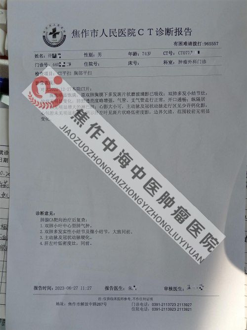
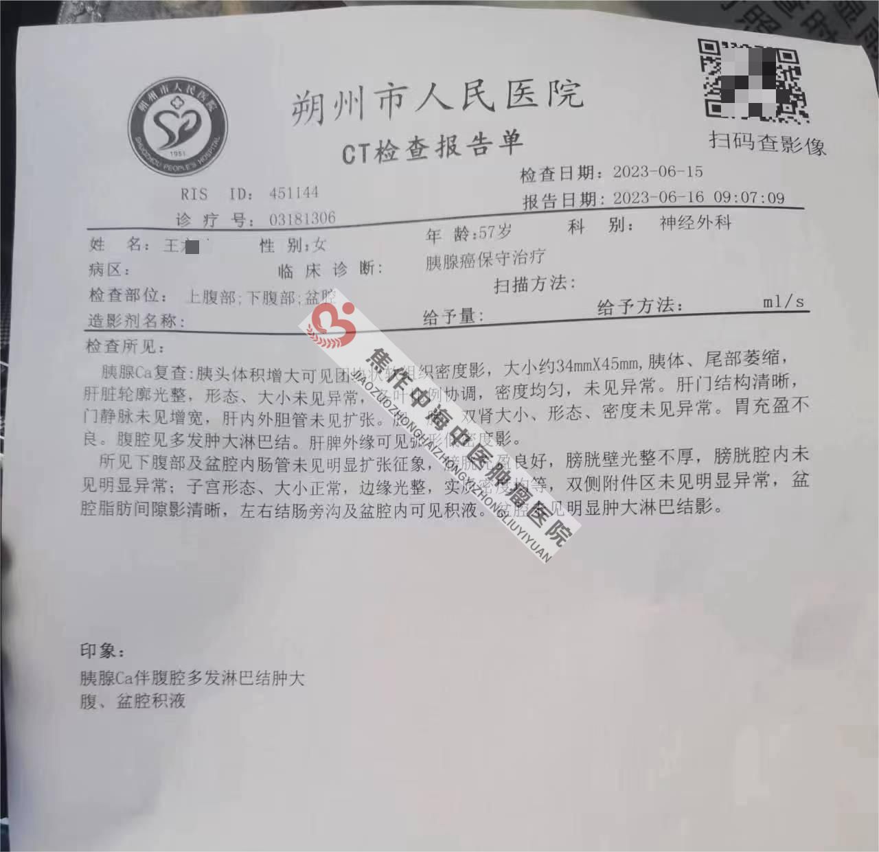
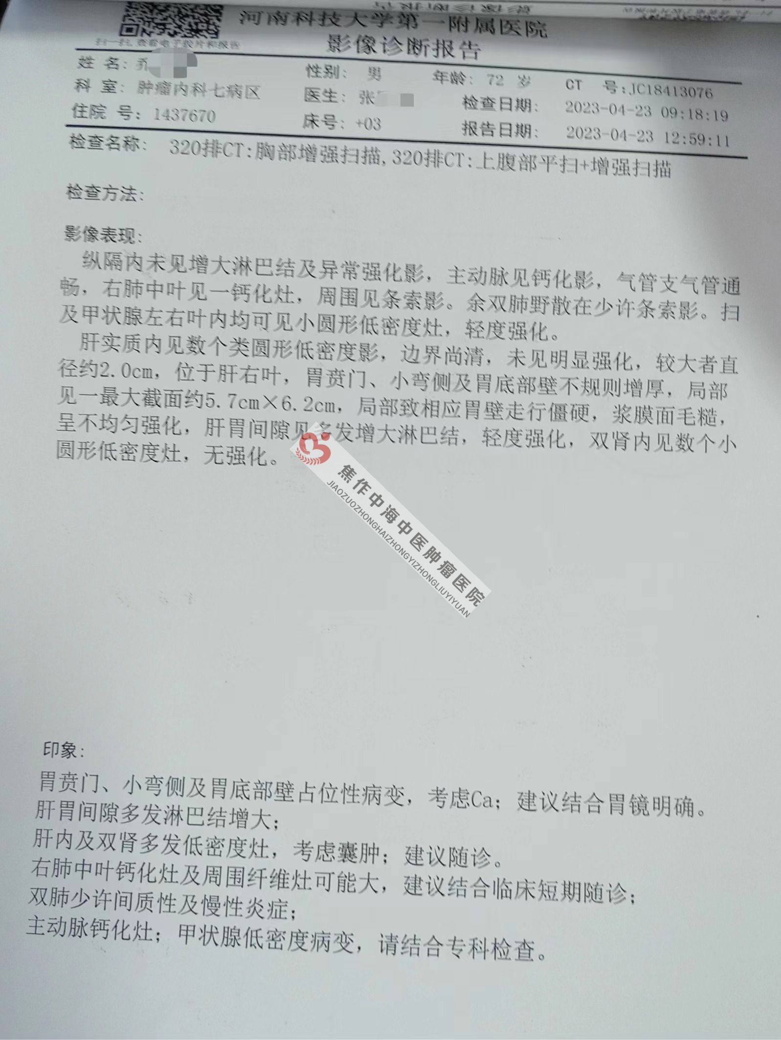
CT examination
CT examination has high resolution and can detect smaller tumors, which has practical value in the diagnosis of liver cancer.
Nuclear magnetic resonance examination
Magnetic resonance imaging (MRI) is a new and valuable diagnostic method for liver cancer, which is approximately 95% qualitatively similar to CT and is also expensive. At present, B-ultrasound examination is generally conducted first, and any problems found can be identified through CT or MRI re examination.