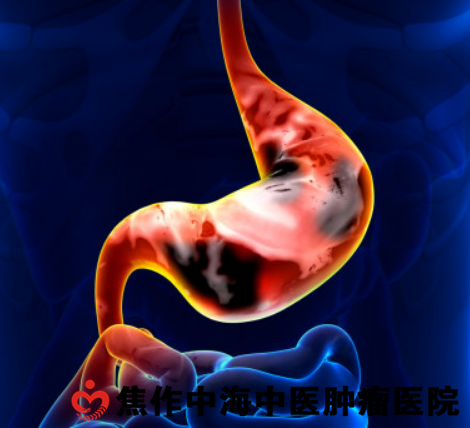
1. Pathology.
(1) Borrmann typing.
Borrmann classification: it is a classic classification method of gastric cancer. This classification is mainly classified according to the morphological characteristics of cancer in the mucous surface and the mode of infiltration in the gastric wall. Gastric cancer is divided into 4 types. Type I (nodular type), the tumor grows into the gastric cavity, the eminence is obvious, the base is wide and the boundary is clear. In type I (ulcer localized type), the tumor had obvious ulcer formation, obvious marginal uplift, and the angle between the base and normal gastric tissue was less than 90 °. Sichuan type (infiltrative ulcerative type), the tumor has obvious ulcer formation, the edge is partially raised, partly infiltrated and destroyed, the boundary is unclear, and the surrounding infiltration is obvious, which is the most common type, accounting for about 50%. Type IV (diffuse infiltrative type) shows diffuse infiltrative growth, so it is difficult to determine the boundary of the tumor. due to the diffuse infiltration of cancer cells and the proliferation of fibrous tissue, the gastric wall thickens and hardens widely. it is called "dermatogastric stomach" WHO put forward the international classification based on tissue origin and atypia in 1979. The system divides gastric cancer into adenocarcinoma (papillary adenocarcinoma, tubular adenocarcinoma, mucinous adenocarcinoma, signet ring cell carcinoma), adenosquamous carcinoma, squamous cell carcinoma, carcinoid, undifferentiated carcinoma and unclassified cancer. When the two types of organizations coexist, they are classified according to the dominant organization, and the secondary organization type is also indicated. According to the degree of differentiation (the least differentiated part), adenocarcinoma can be divided into highly differentiated type, moderately differentiated type and poorly differentiated adenocarcinoma. In 1990, WHO revised the histological classification of gastric cancer. According to the new standard, gastric cancer was divided into epithelial tumors and carcinoid tumors. Epithelial tumors included adenocarcinoma (papillary adenocarcinoma, tubular adenocarcinoma, poorly differentiated adenocarcinoma, mucinous adenocarcinoma, signet ring cell carcinoma), squamous adenocarcinoma, unpulverized carcinoma and unclassified carcinoma. The microscopic structures of various tissue types of tumors are different: 1 Tubular adenocarcinoma: the cancer tissue shows glandular tube-like or acinar structure. According to the degree of cell differentiation, it can be divided into two types: high and middle differentiation. 2 papillary adenocarcinoma: the cancer cells were generally well differentiated, cuboidal or high columnar, arranged around the slender dendritic stroma to form papillary structures of different thickness. (3) poorly differentiated adenocarcinoma: the cancer cells were short columnar or amorphous, arranged in the shape of small nest or cord, and basically had no glandular tube structure. According to the number of stroma, it can be divided into solid type and non-solid type. (4) mucinous adenocarcinoma: it is characterized by cancer cells forming lumens and secreting a large amount of mucus. Due to the accumulation of a large amount of mucus, many glandular cavities are expanded or ruptured, and mucus infiltrates the stroma, that is, to form a "mucus lake". 5 signet ring cell carcinoma: secretes a large amount of mucus for cancer cells, and the mucus is located in the cell, pushing the nucleus to the periphery of one side of the cell, and the whole cell is in the shape of a signet ring. Its malignant degree is higher than that of extracellular mucus. 6 adenosquamous carcinoma: also known as glandular spinous cell carcinoma, is a kind of tumor with the coexistence of adenocarcinoma and squamous cell carcinoma. Some cells of adenocarcinoma are well differentiated, while some cells of squamous cell carcinoma are poorly differentiated. (7) squamous cell carcinoma: most of the cells were moderately to poorly differentiated, showing a typical squamous cell carcinoma structure, and those involving the end of the esophagus should be considered to be caused by the expansion of primary esophageal squamous cell carcinoma. 8 undifferentiated cancer: cancer cells disperse into patches or lumps.Shape, do not form a tubular structure or other organizational structure. The cells are small in size, obvious in heteromorphism, and lack of differentiation characteristics in tissue morphology and function. 9 carcinoid: a low-grade malignant tumor from the argyrophil cells at the bottom of the digestive tract gland. the cancer cells are small, uniform and densely arranged, and dark-brown argyrophil granules can be seen in the cytoplasm by argyrophil staining.2. Metastatic pathway of gastric cancer.1. The cancer tissue directly infiltrating into the gastric serosa can spread directly to the adjacent organs and tissues, such as the liver, pancreas and greater omentum.

